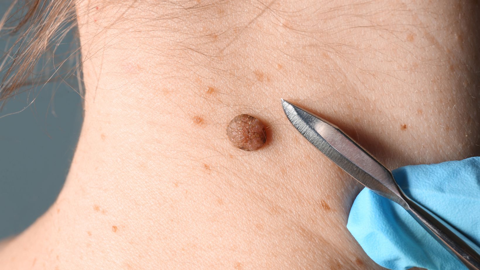
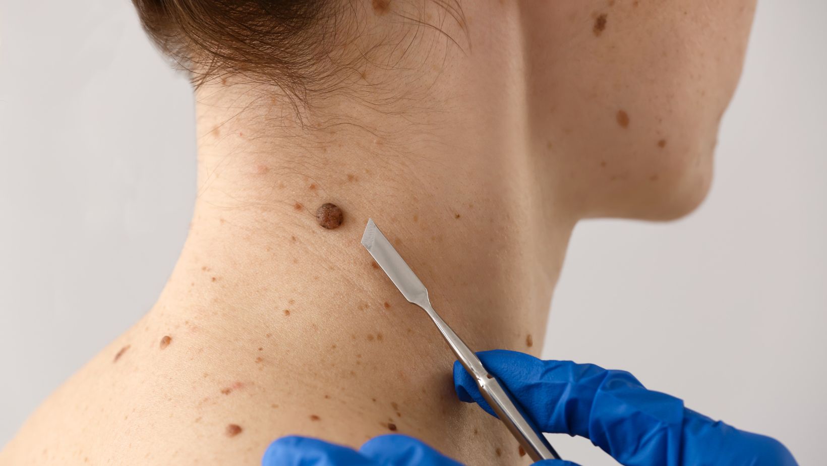
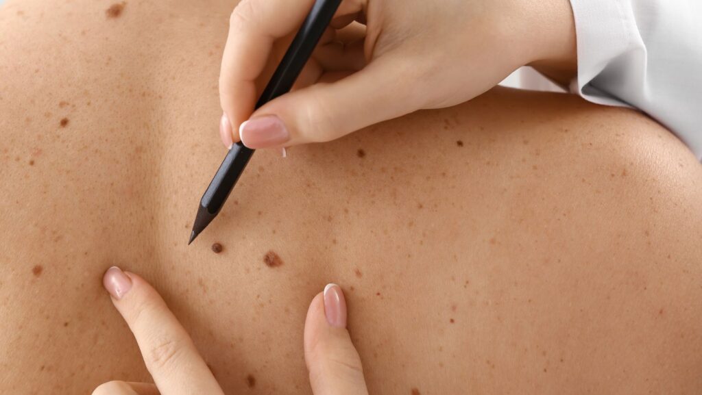
Moles are common skin growths that most people have. While many moles are harmless, some can be a sign of skin cancer. It’s important to check moles regularly for changes in size, shape, or color.
Dermatologists can test suspicious moles by taking a small sample and examining it under a microscope. This simple procedure can detect cancer cells if present. Knowing the signs of a cancerous mole can help people seek medical care early when treatment is most effective.
There are several ways to safely remove moles. Options include laser treatments and surgical excision by a dermatologist. The best method depends on the mole’s size and location. Removal can improve appearance and ease worries about cancer risk.
Understanding Moles and Skin Health
Moles are common skin growths that can appear anywhere on the body. Most moles are harmless, but some may develop into skin cancer. Knowing the difference between normal and suspicious moles is key for skin health.
Characteristics of Benign vs. Cancerous Moles
Normal moles are usually round or oval with smooth edges. They have an even color and stay the same size over time. Cancerous moles look different.
The ABCDE rule helps spot warning signs:
- A: Asymmetry (uneven shape)
- B: Border (jagged or blurry edges)
- C: Color (multiple shades or changes)
- D: Diameter (larger than 6mm)
- E: Evolving (changes in size, shape, or color)
Atypical moles may have some of these features but aren’t cancer. A dermatologist can check any moles that worry you.
Prevention and Early Detection Strategies
Protecting your skin from the sun is the best way to prevent skin cancer. Use sunscreen, wear protective clothing, and avoid tanning beds.
Skin self-checks are important. Look at your skin once a month and note any new or changing moles. People with fair skin, many moles, or a family history of melanoma have a higher risk.
See a doctor for regular skin cancer screenings. They can spot issues early when treatment works best. Make an appointment right away if you notice any moles with ABCDE warning signs.
Diagnostic Procedures for Moles
Doctors use specific tests to check if moles are cancerous. These tests help find any signs of skin cancer early.
Biopsy Techniques and Analysis
A skin biopsy is the main way to test moles. A dermatologist takes a small piece of the mole to look at under a microscope. There are different types of biopsies.
Shave biopsies remove the top layers of skin. Punch biopsies go deeper and take out a small round piece. Excisional biopsies cut out the whole mole and some skin around it.
After the biopsy, a lab checks the sample.

They look for cancer cells or weird cell growth. The results help decide if more treatment is needed.
Before a biopsy, doctors check moles closely. They look for things like odd shapes, colors, or sizes. These “ugly ducklings” may need testing.
Most moles are harmless. But it’s good to have a doctor check any that seem strange.
Mole Removal Techniques and Post-Procedure Care
Mole removal is a common procedure that can be done for cosmetic or medical reasons. There are several methods available, ranging from surgical to non-invasive options. Proper aftercare is key to ensuring good healing and minimizing scarring.
Surgical and Non-Surgical Removal Options
Doctors use different techniques to remove moles based on their size, location, and type. Surgical excision involves cutting out the mole and some surrounding skin. This method works well for larger or potentially cancerous moles.
Shaving is another option where the mole is sliced off at skin level. It’s best for smaller, raised moles. Cryotherapy uses liquid nitrogen to freeze the mole, causing it to fall off. This works well for some types of benign moles.
Electric current can burn off moles in a process called electrocautery. It’s quick and leaves little scarring. For shallow moles, doctors might use a scalpel to scrape them off the skin’s surface.
Laser Treatment Methods for Mole Removal
Laser treatments offer a non-surgical way to remove moles. The Ultrapulse CO2 laser is good for deeper moles. It uses very short pulses of light to remove layers of skin.
Pigment lasers target the color in moles. They work best on dark, flat moles. These lasers break up the pigment, which the body then absorbs.
Laser treatments often need multiple sessions. They can be less painful than surgery and may leave less scarring. But they might not work as well on raised moles.
Aftercare and Monitoring
After mole removal, proper care helps prevent infection and reduce scarring. Keep the area clean and dry.

Apply petroleum jelly to the wound and cover it with a bandage.
Avoid picking at the scab or exposing the area to sunlight. Watch for signs of infection like redness, swelling, or pus. Call your doctor if you notice these signs.
For some removal methods, you might need follow-up visits. Your doctor may want to check how the site is healing. They may also test the removed mole for cancer cells.
It’s important to keep an eye on your skin after mole removal. New moles can still form. Check your skin regularly for any changes. See a doctor if you notice any new or changing spots.
